Atlas Color
Cross-Sectional Atlas of the Human Head: With 0.1-mm pixel size color images (Repost) eBooks & eLearning
Posted by AvaxGenius at Jan. 5, 2025
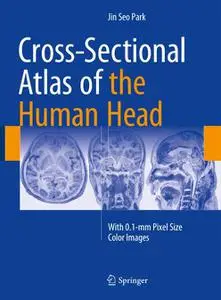
Cross-Sectional Atlas of the Human Head: With 0.1-mm pixel size color images By Jin Seo Park
English | PDF,EPUB | 2017 | 328 Pages | ISBN : 9811007691 | 159.64 MB
This superb color atlas sets a new standard in neuroanatomy by presenting around 300 detailed thin-sectioned images of the human head, including the brain, with 0.1-mm intervals and a pixel size of 0.1 mm × 0.1 mm. A new reference system employed for this purpose is clearly explained, and structures are fully annotated in the horizontal, coronal, and sagittal planes. Recent advances in 7T MRI and 7T TDI have considerably enhanced imaging of the human brain, thereby impacting on both neuroscience research and clinical practice. Moreover, the information gained from initiatives involving photography of thin slices of human cadavers, such as the Visible Human Projects, Visible Korean and Chinese Visible Human, has enriched knowledge of neuroanatomy and thereby facilitated the interpretation of such ultra-high-field resolution images. The exquisite images contained within this atlas will be invaluable in providing both researchers and clinicians with important new insights.
Atlas of Fine Needle Aspiration Cytology (Repost) eBooks & eLearning
Posted by AvaxGenius at July 9, 2022
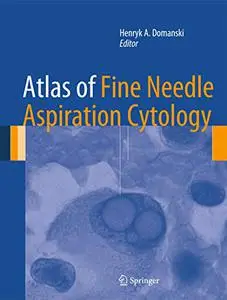
Atlas of Fine Needle Aspiration Cytology by Henryk A. Domanski
English | PDF(True) | 2014 | 572 Pages | ISBN : 1447124456 | 120 MB
This book covers all of the diagnostic areas where FNAC is used today. This includes palpable lesions and lesions sampled using various radiological methods, and correlations with ancillary examinations detailed on an entity-by-entity basis. As well as being a complete atlas of the facts and findings important to FNAC, this atlas is a guide to diagnostic methods that optimize health care. The interaction of the cytologist or cytopathologist with other specialists (radiologists, oncologists and surgeons) involved in the diagnosis and treatment of patients with suspicious mass lesions is emphasized and illustrated throughout.
Clinical Atlas of CT Virtual Hysterosalpingography (Repost) eBooks & eLearning
Posted by AvaxGenius at July 1, 2022
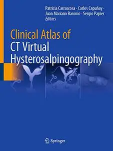
Clinical Atlas of CT Virtual Hysterosalpingography edited by Patricia Carrascosa
English | EPUB | 2021 | 419 Pages | ISBN : 3030662063 | 458.8 MB
This book provides a comprehensive, practically applicable guide to the use of CT virtual hysterosalpingography for evaluating gynaecological pathology and infertility in women. It features detailed descriptions of normal and pathologic findings across the female reproductive system, including the cervix, uterine wall and cavity, and Fallopian tubes, and compares the findings with other imaging modalities such as ultrasound, X-ray hysterosalpingography and MRI. The interpretation of post-treatment findings and commonly encountered pitfalls are also covered in detail.
Comprehensive Atlas of Dermatoscopy Cases eBooks & eLearning
Posted by AvaxGenius at June 27, 2022
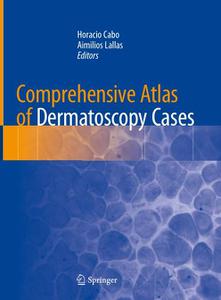
Comprehensive Atlas of Dermatoscopy Cases by Horacio Cabo
English | EPUB | 2018 | 215 Pages | ISBN : 3319769316 | 125.9 MB
This practical atlas describes the use of dermoscopy in the clinic, a technique that is increasingly used by the clinical dermatologist. It revolves around the use of clinical cases, simulating what happens in the clinic when the dermatologist is presented with a patient who has pigmented lesions. Dermatologists perform diagnoses based on what they see on the skin and with these images recognize different diseases. This whole spectrum of forms and shapes is reflected in colour.
Color Atlas of Spinal Cord Surgery eBooks & eLearning
Posted by IrGens at July 8, 2022

Color Atlas of Spinal Cord Surgery by Jörg Klekamp
English | May 21, 2022 | ISBN: 3030899632 | True EPUB | 541 pages | 1.05 GB
English | May 21, 2022 | ISBN: 3030899632 | True EPUB | 541 pages | 1.05 GB
Color Atlas of Forensic Medicine and Pathology, 2nd Edition eBooks & eLearning
Posted by IrGens at Nov. 27, 2022

Color Atlas of Forensic Medicine and Pathology, 2nd Edition edited by Charles Catanese
English | March 2, 2016 | ISBN: 1466585900 | True EPUB | 634 pages | 548 MB
English | March 2, 2016 | ISBN: 1466585900 | True EPUB | 634 pages | 548 MB
Emily Bloom by Atlas Elison Girls
Posted by nrg at Feb. 18, 2022

Color Atlas of Medical Bacteriology Ed 3 eBooks & eLearning
Posted by arundhati at May 16, 2020
Luis M. de la Maza, "Color Atlas of Medical Bacteriology Ed 3"
English | ISBN: 1683670353 | 2020 | 464 pages | PDF | 51 MB
English | ISBN: 1683670353 | 2020 | 464 pages | PDF | 51 MB
The SAGES Atlas of Robotic Surgery (Repost) eBooks & eLearning
Posted by AvaxGenius at April 20, 2021

The SAGES Atlas of Robotic Surgery by Yuman Fong
English | PDF,EPUB | 2018 | 502 Pages | ISBN : 3319910434 | 622.66 MB
This book is intended as a definitive, state of the art guide to robotic surgery that summarizes the field for surgeons at all levels. More specifically, its goals are threefold: to review the basics of robotic surgery, including fundamental principles, technology, operating room setup, and workflow; to describe and illustrate the procedures most commonly performed in a robotic operating room; and to discuss key issues relating to cost, adoption, and training.
Atlas of Deep Endometriosis: MRI and Laparoscopic Correlations (Repost) eBooks & eLearning
Posted by AvaxGenius at May 2, 2021
Atlas of Deep Endometriosis: MRI and Laparoscopic Correlations By Alice Brandão
English | PDF,EPUB | 2018 | 368 Pages | ISBN : 3319716964 | 146.21 MB
This Atlas presents an MRI-based guide to the diagnosis, treatment and follow up of deep endometriosis. Developed by professionals with a extensive clinical experience in the diagnosis and treatment of deep endometriosis, it provides a global overview of the disease, from basic clinical aspects of imaging diagnosis, to the correlation with surgical findings and histopathological results.